There are various types of tissue.
Epithelial Tissue:
Simple Squamous- this photo shows Alveoli of the lungs
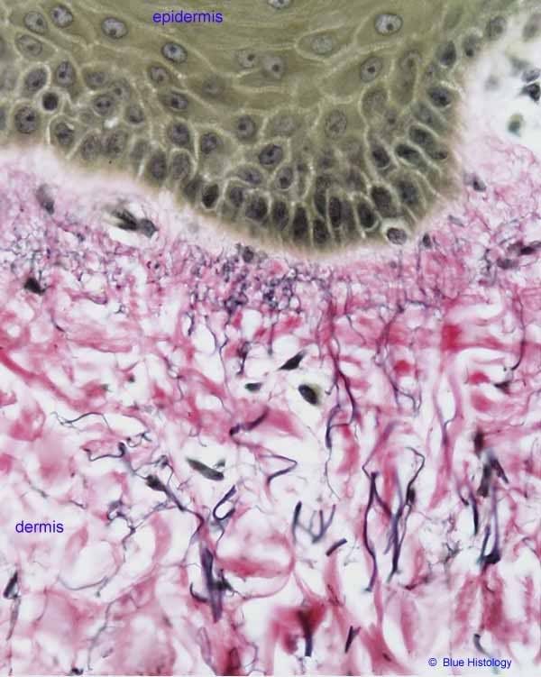
Stratified Squamous- Keratinized in skin
Pseudostratified Ciliated Columnar Epithelium
Simple Cuboidal Epithelium-
Simple Columnar Epithelium-
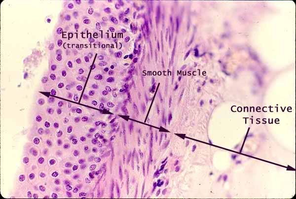
Transitional Epithelium-
CT- Connective Tissue
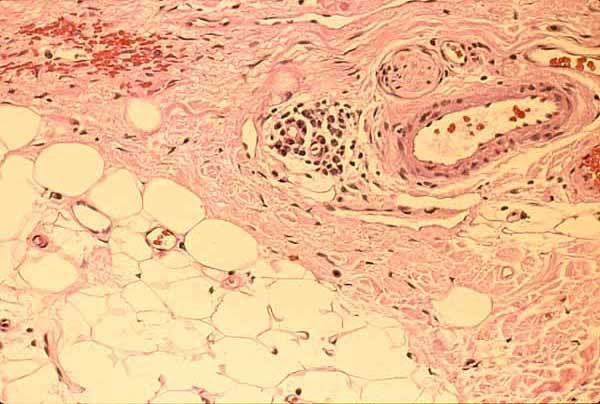
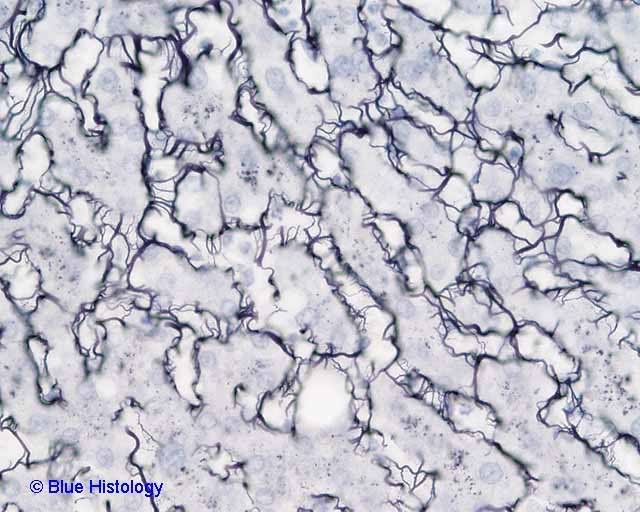
Areolar CT
This image of areolar CT represents a whole mount (NOT a section) of mesentery (the tissue which binds together the loops of intestine within the peritoneal cavity.
This view looks through the entire mesentery, which is a sandwich of mesothelium on front and back surfaces with loose connective tissue and blood vessels in between.
The two most common cell types are mesothelial cells and fibroblasts.
The large, pale, oval nuclei belong to mesothelial cells which form the mesothelial surfaces on the front and back of the mesentery.
The smaller, darker, more elongate nuclei belong to fibroblasts, which manufacture and secrete the molecules which form the extracellular matrix.
Macrophages, mast cells, and lymphocytes may also be found in such preparations, but none are clearly identifiable in this image.
The matrix consists of unstained ground substance through which pass fibers of made of collagen and elastin.
Elastic fibers are thin, fairly uniform in diameter, and frequently branched. The are normally visible only if specially stained (as they have been in this specimen).
Collagen fibers vary in thickness and are eosinophilic (pink, in this stain).
CT- Elastic Fibers
CT- Reticular CT silver stain only
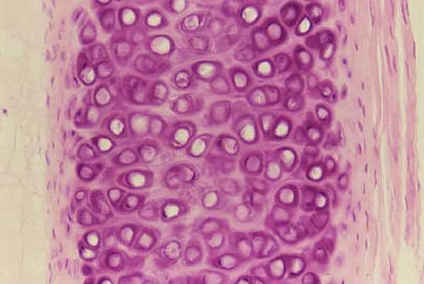
CT- Hyaline Cartilage Chondrocytes in Lacunae
CT- Adipose fat storage